
Case vignette
56yo M w/ DM2 p/w progressive R foot swelling and pain. It started a few days ago but got a lot worse today.
HR 115, BP 110/79, T 37.7, RR 19, SpO2 96%
R foot has erythema and swelling over dorsum extending towards distal leg. Foot is very tender to touch and feels tense.
What do you see?
MSK3 US1: cobblestoning consistent with edema/inflammation. This transitions to hyperechoic artifact with shadowing beneath it
MSK3 US2: re-demonstrated hyperechoic artifact with posterior shadowing
What do you do next?
You recognize this hyperechoic artifact/shadowing is a very, very bad sign: gas. You place an emergent call to Gen Surg and order a septic work up and CK. You order Clindamycin first, followed by Zosyn and Linezolid. You also order morphine. Gen Surg arrives, agrees the patient has signs of necrotizing fasciitis, and rushes the patient to the OR for debridement.
The patient has a complex ensuing course requiring BKA but ultimatley survives.

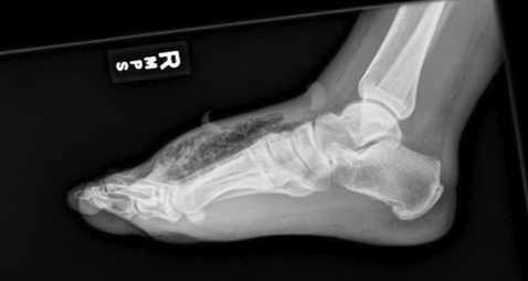
The below XR was obtained in the ED too, by the way, and read the next day by radiology as concerning for gas.
Teaching points. Necrotizing soft tissue infections (NSTIs) are rare but devastating. Morbidity and mortality increases substantially with each passing hour. NSTIs are also notoriously difficult to catch. While risk stratification tools such as LRINEC exists, no single test or tool is reliable. Even CT is not definitive: remember, this is a clinical diagnosis. Ultimately, the only way to prove or disprove NSTI is with a knife in the fascial tissue.
History and exam may demonstrate pain out of proportion, rapidly progressive cellulitic changes, dark discoloration or bullae, crepitus, systemic symptoms such as fever or N/V/D, and immunosuppression. That said, many of these findings may be absent or occur late. Consider imaging to look for gas, which would increase your pre-test probability. US, XR, and CT can be used. On US, gas typically appears as a bright (hyperechoic) area with shadowing beneath it. While this has low sensitivity and does not occur with all NSTIs, it is better than XR and immediately available. CT is more sensitive and can direct surgical efforts but does take more time.
Concern for this diagnosis should prompt immediate Surgical consult, septic work up, and broad spectrum antibiotics. Three antibiotics are typically used. Clindamycin, often given first, reduces toxin exposure for some microorganisms (e.g., Group-A Strep). Second is typically a broad-spectrum beta-lactam such as Zosyn. Finally, MRSA coverage may be best with Linezolid. Linezolid may outperform Vancomycin by also minimizing toxin exposure. Additional treatments being studied include IVIG, for example for concurrent toxic shock, and hyperbaric oxygen.
Sources: Farkas J. Necrotizing fasciitis. EmCrit: The Internet Book of Critical Care. March 2021. https://emcrit.org/ibcc/necfas/. Accessed 4/20/21.
Residents: Dr. Sam Ho
Necrotizing Soft Tissue Infection
July 2018