

Case Vignette
A 53-year-old man with atrial fibrillation and a history of unprovoked deep venous thrombosis on rivaroxaban presented to the Emergency Department with new flashes and floaters in the right eye.
Four weeks earlier, he had been struck in the right eye by a foam toy projectile from approximately 20 feet away. He experienced minimal pain and initially noticed several small dark spots in his vision, which resolved after about two days.
On the day of presentation, he awoke with gold, ring-like flashes in the right eye, followed by a larger dark floater in the temporal visual field. The flashes later resolved, but the floater persisted. He denied eye pain, fixed visual field loss, complete vision loss, headache, or focal neurologic symptoms.
Visual acuity was 20/25+1 bilaterally, intraocular pressures were 10 mmHg in the right eye and 12 mmHg in the left, and pupils were briskly reactive. Visual fields and extraocular movements were normal.
Bedside ultrasound was performed as shown below:
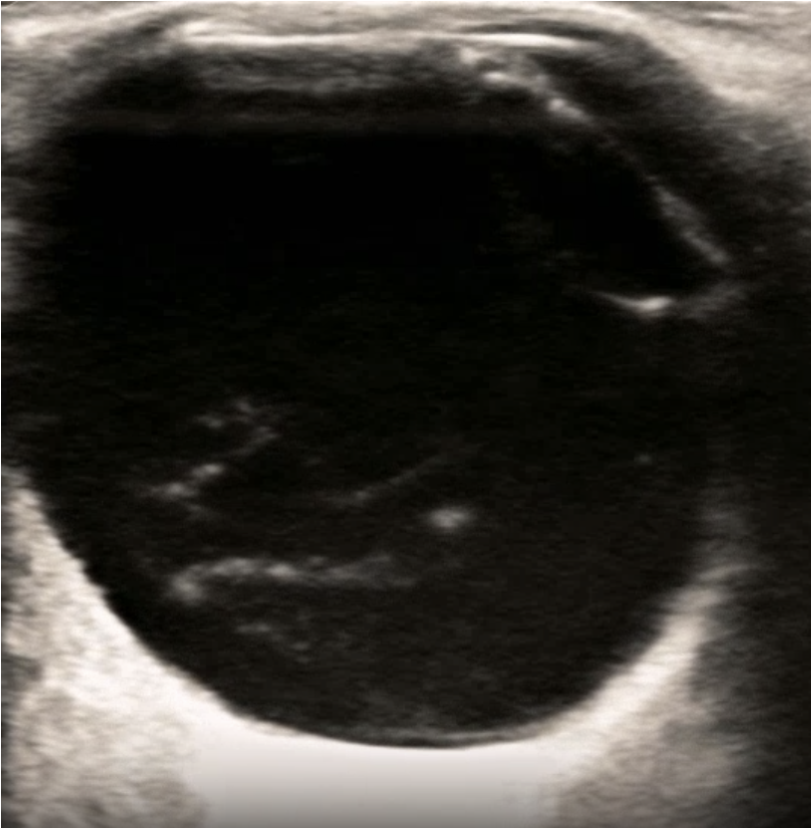
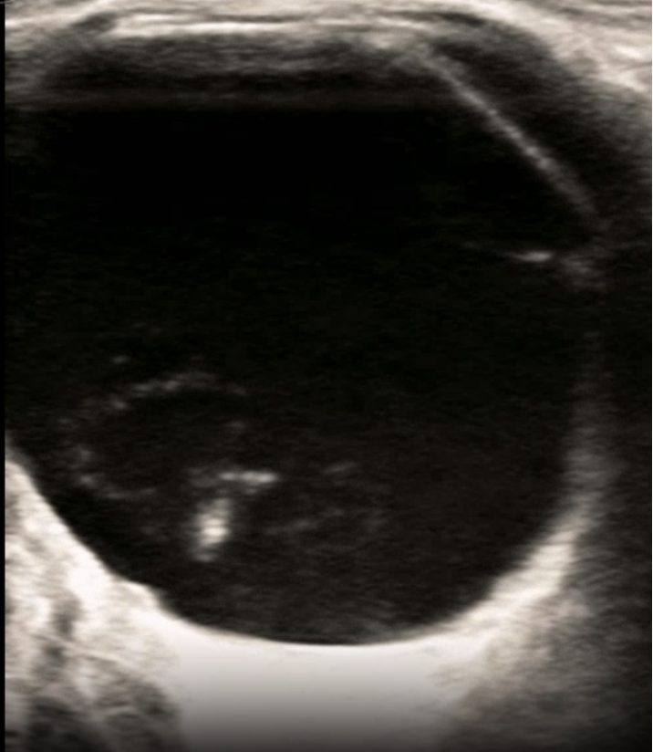
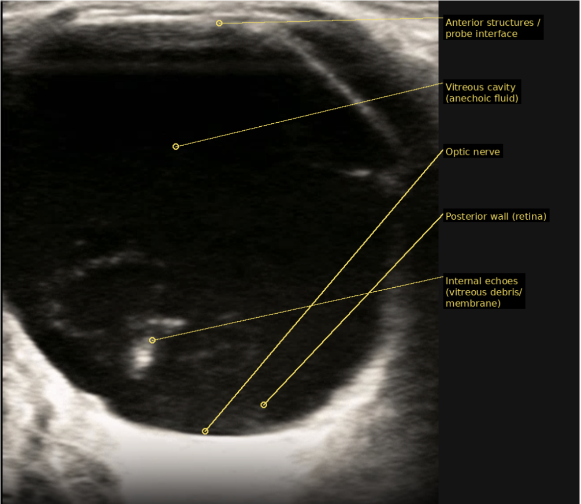
Ophthalmic ultrasound in the Emergency Department showed a thin echogenic membrane within the posterior vitreous cavity, separated from the retinal surface, consistent with posterior vitreous detachment, which is shown in the images above.
He was diagnosed with symptomatic posterior vitreous detachment of the right eye and ophthalmology consulted to establish close follow up. The dilated ophthalmologic examination showed a Weiss ring in the right eye with a negative Shafer sign and an attached retina on 360-degree scleral depressed examination. They agreed with the diagnosis and established follow-up within one week.
Intro:
PVD is the separation of the posterior vitreous cortex from the retina's internal limiting membrane and is often an age-related process(1). Prevalence rises significantly with age, from roughly 24% in patients aged 50–59 years to 87% in those aged 80–89 years(2). PVD typically often occurs more frequently and earlier in men, those with myopia, and after trauma or intraocular surgery(1).
Trauma is a recognized precipitant of earlier-onset PVD, relevant to this patient whose symptoms followed blunt injury and who lacked other classic risk factors such as high myopia or prior surgery(3). As the vitreous separates and pulls at sites of vitreoretinal adhesion, this traction can cause full-thickness retinal breaks, allowing liquefied vitreous to enter the subretinal space and cause retinal detachment(1).
Classic symptoms include photopsias (light flashes, often most noticeable in the dark) and floaters (myodesopsias). Floaters may represent condensed vitreous collagen, blood from a torn retinal vessel, or glial tissue seen clinically as a Weiss ring, which is the hallmark of posterior vitreous separation identified in this patient(1). PVD may be asymptomatic in up to 20% of cases(3).
Discussion:
PVD is typically an age-related event in which the posterior vitreous cortex separates from the retina's internal membrane. Blunt ocular trauma is a well-recognized accelerant of this process. Trauma-inducedalterations of the vitreoretinal interface may manifest either at the time of injury or years later(1). Posterior-segment trauma can cause vitreous separation, vitreous hemorrhage, and retinal tears(4).
For this patient, the four-week interval between blunt ocular impact and the onset of photopsias and floaters is consistent with a trauma-accelerated PVD. The transient dark spots the patient noted immediately after injury may have reflected early vitreous disturbance, while the subsequent gold ring-like photopsias and new temporal floater represented completion of posterior vitreous separation, ultimately confirmed by a Weiss ring on dilated examination. The flashes reflect vitreoretinal traction during active separation; as the vitreous finishes detaching, the traction, along with the flashes, abate.
The central clinical concern in acute symptomatic PVD is identifying a concurrent or subsequent retinal break. In a large Kaiser Permanente cohort of acute PVD, the presence of vitreous hemorrhage, vitreous pigment (a positive Shafer sign), or lattice degeneration markedly elevated the risk of retinal tear or detachment (12.4% risk), whereas eyes lacking all of these features carried a low late-complication risk of 0.7%. This patient had a negative Shafer sign, no vitreous hemorrhage, no lattice degeneration, and an attached retina without an identifiable break, placing him in the low-risk category for immediate complications. PVD (without retinal break) symptoms usually diminish over time, sometimes requiring several months. Improvement occurs partly through neuroadaptation and partly because the Weiss ring and vitreous opacities migrate anteriorly and peripherally, away from the visual axis(1).
Conclusion:
Delayed symptomatic posterior vitreous detachment may occur after seemingly minor blunt ocular trauma. New flashes or floaters after eye injury should prompt evaluation for associated retinal tears or detachment, even when visual acuity is preserved and the external examination is reassuring.
References:
Kim S, et al. Posterior Vitreous Detachment, Retinal Breaks, and Lattice Degeneration Preferred Practice Pattern® Ophthalmology, 2025; 132, P163-P196
Hollands H, Johnson D, Brox AC, Almeida D, Simel DL, Sharma S. Acute-Onset Floaters and
Flashes: Is This Patient at Risk for Retinal Detachment? JAMA. 2009;302(20):2243–2249.
doi:10.1001/jama.2009.1714
Driban, M., Chhablani, J. Clinical findings in acute posterior vitreous detachment. Graefes Arch
Clin Exp Ophthalmol 260, 3465–3469 (2022). https://doi.org/10.1007/s00417-022-05708-4
Williams DF, Mieler WF, Williams GA. Posterior segment manifestations of ocular trauma.
Retina. 1990;10 Suppl 1:S35-44. doi: 10.1097/00006982-199010001-00006. PMID: 2191381.
Seider MI, Conell C, Melles RB. Complications of Acute Posterior Vitreous Detachment.
Ophthalmology. 2022 Jan;129(1):67-72. doi: 10.1016/j.ophtha.2021.07.020. Epub 2021 Jul 27.
PMID: 34324945.
Lily Sloan
Vitreous Detachement
9/2026