

Case Vignette
77-year-old male with past medical history of BPH, hypertension, type 2 diabetes and chronic encephalopathy presented to emergency department referred by his skilled nursing facility (SNF) with chief complaint of altered mental status. Prior to presentation, patient was found by nursing staff at his SNF to have decreased responsiveness. EMS was called and patient was brought to OHSU ED. En route to the emergency department, patient was afebrile with blood pressure 107/54, HR 88, RR 18, with SpO2 97% on RA. CBG 176. Patient had an episode of non-bloody, non-bilious emesis in the ambulance and was given 8mg IV Zofran.
Initial ED course:
Upon presentation to the ED, patient reported fever, abdominal pain, and difficulty urinating, unsure of the last time he voided. Patient presented alert and oriented x 2-3 to self and location, consistent with his baseline mental status per family. Physical exam was notable for soft, tender abdomen with mild distention in suprapubic region. Initial differential diagnosis of emergency medicine physician included urinary tract infection with urinary retention, metabolic disturbance, and hydronephrosis. Initial labs demonstrated Na 128, K+ of 6.3, Cr of 3.72 (1.2 baseline), and BUN 100. CBC was negative for leukocytosis, demonstrated stable anemia. Urinalysis demonstrated WBCs>1000, RBC>1000, many bacteria, leukocyte esterase positive, and nitrite negative, suggest of urinary tract infection. EKG demonstrated NSR with peaked T waves. PVR 377mL. Patient was given Lokelma, insulin regular, 50mL D50, and calcium gluconate for his hyperkalemia.
POCUS:
Bedside POCUS of patient’s bladder and kidneys was performed to evaluate for hydronephrosis given concern for urinary retention and findings of AKI on lab work. Renal and bladder views were obtained using curvilinear probe. The kidneys were scanned through in two axes, longitudinal and transverse. Scan of patient’s right kidney (Figure 1) demonstrated pelviectasis and caliectasis, dilatation of the renal pelvis and calyces, respectively. This gross dilatation appeared to result in some cortical thinning, consistent with moderate-severe grade hydronephrosis.1 Bedside ultrasound of patient’s left kidney (Figure 2) demonstrated a dilated renal pelvis and enlarged calyces and hydroureter were visualized as well. Cortical thinning was not as apparent on the scans of the left kidney as compared to the right. Findings were consistent with moderate-severe grade hydronephrosis.
Scan of the bladder demonstrated an indwelling foley catheter and heterogenous layering fluid, thought possibly to be blood given patients microscopic hematuria on UA (Figure 3). Volume was calculated to be approximately 497mL using the correction coefficient of 0.7 (9.52cm *8.29cm*~9cm*0.7), suggestive of bladder distention and urinary retention.2 The prostate with inserted foley catheter was visualized on longitudinal view of the bladder and appeared grossly enlarged, suggestive as cause of patient’s obstructive uropathy (Figure 4).
Color doppler was also placed over the renal pelvis (Figure 5) and trigone of the bladder (Figure 6). Color was placed on the renal pelvis and trigone of bladder near the uterovesical junction to evaluate for twinkling artifact which would suggest presence of obstructing renal or ureteral stone. No multicolored comet-like signal that would suggest turbulent flow behind an obstructing stone was visualized. Additionally, no ureteral jets were visualized at the level of the trigone of the bladder that would represent urine flow from kidney to bladder. This is usually evaluated when obstructive ureteral stone is suspected and presence of unilateral jet can suggest obstruction on contralateral side. However, given intermittent release of urine from kidney to bladder, jets can occur as infrequently as 10 minutes and it is recommended to evaluate for up to 10 minutes to confirm absence of jets which was not done in this case. Therefore, absence of jets bilaterally in this situation did not increase suspicion for bilateral renal stones given the enlarged prostate was the more likely etiology of bilateral hydronephrosis.
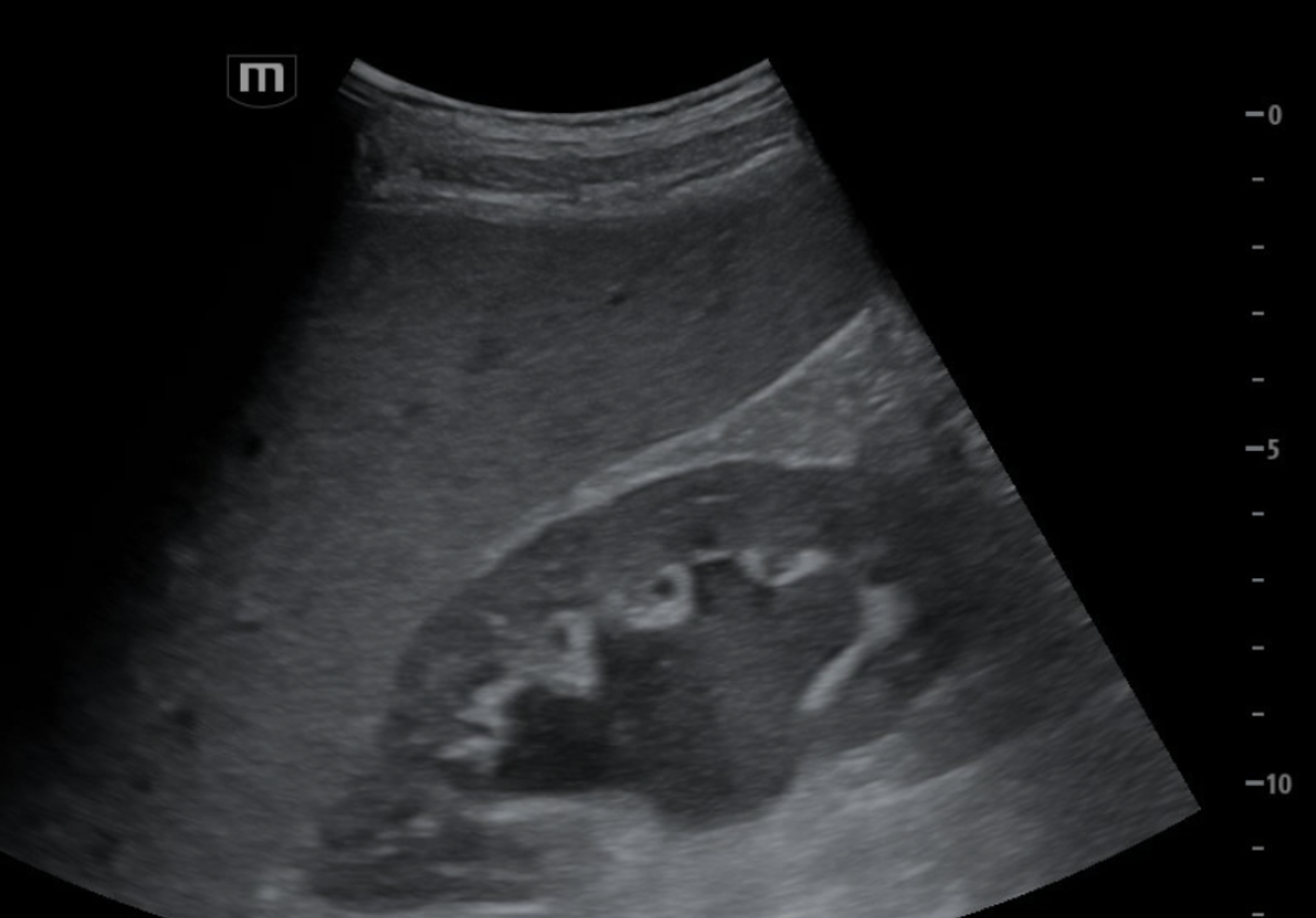
Figure 1: Patient’s right kidney in longitudinal view with renal pelvis, major calyces, and minor calyces visualized. Some cortical thinning present as well.
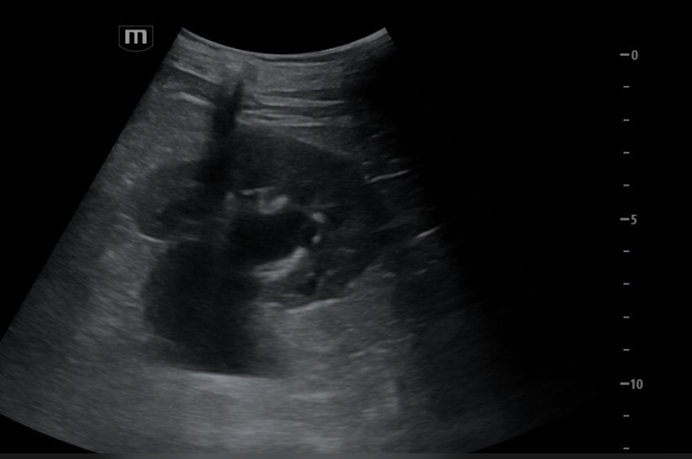
Figure 2: POCUS of left kidney in longitudinal view, demonstrating pelviectasis and caliectasis. Hydroureter inferior to the kidney seen as well.
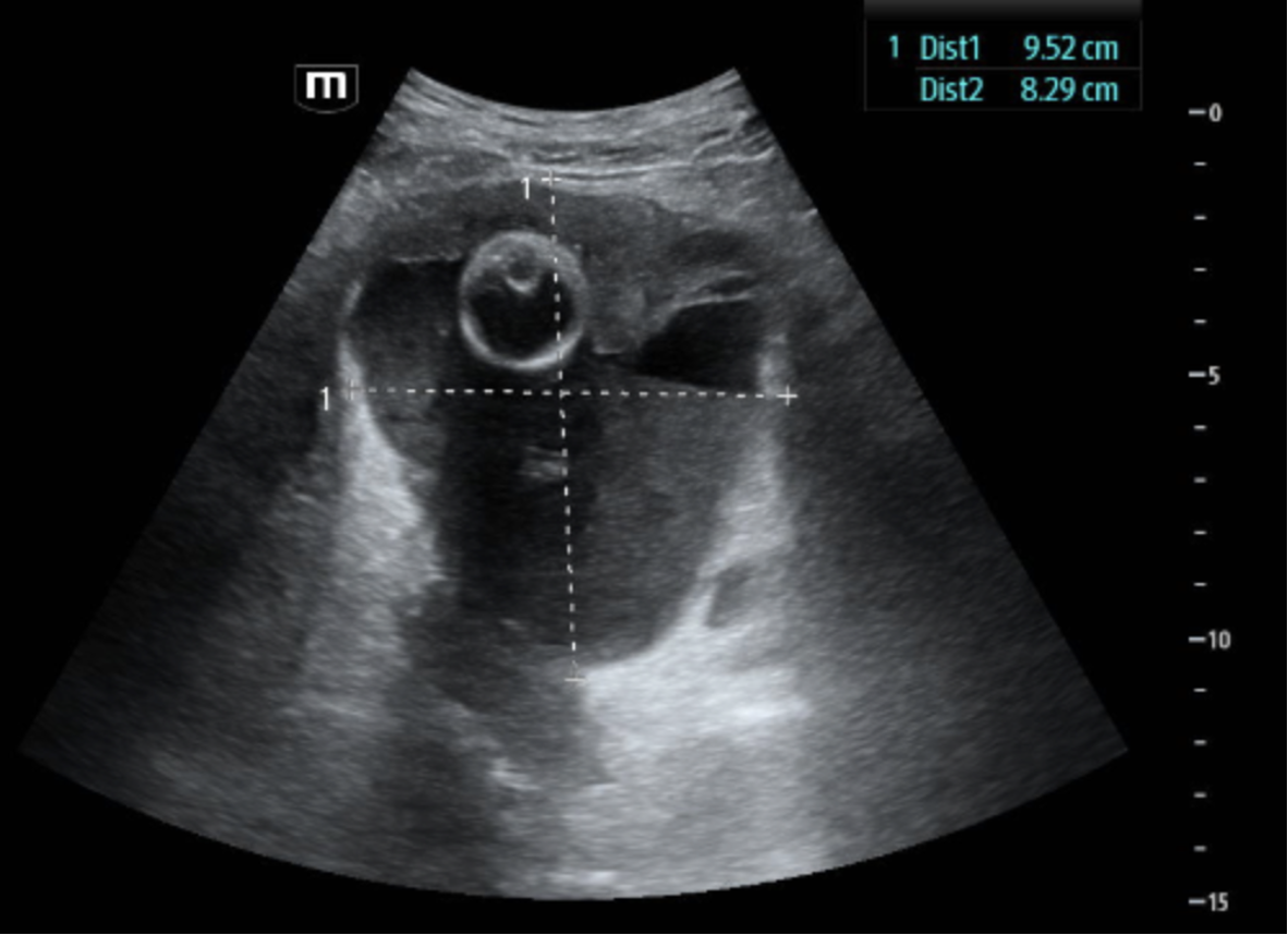
Figure 3: Transverse view of bladder with heterogenous fluid and foley catheter in place. Height and width measurements marked.
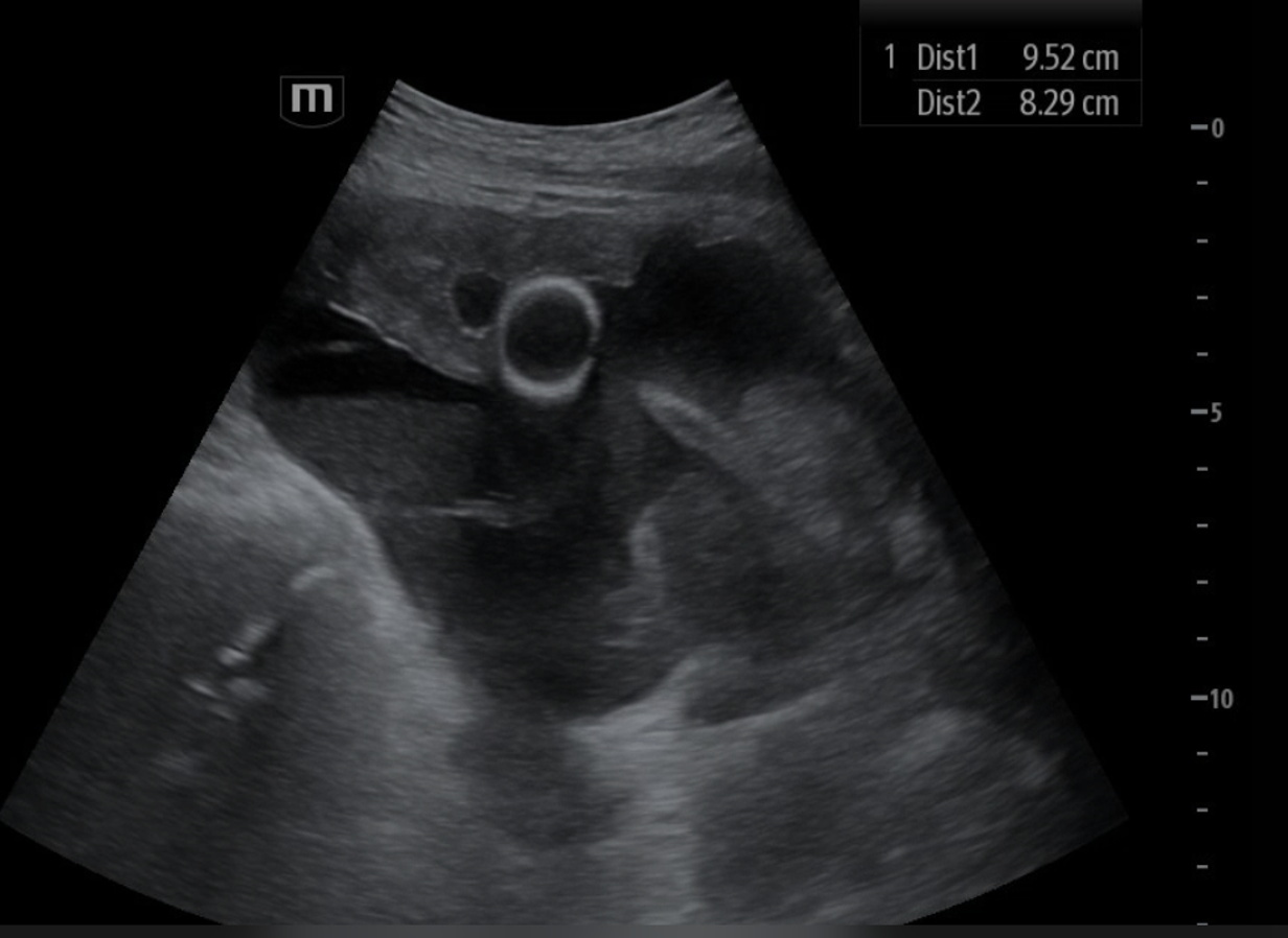
Figure 4: Longitudinal view of bladder with grossly appearing enlarged prostate inferior to bladder and on right side of scan.
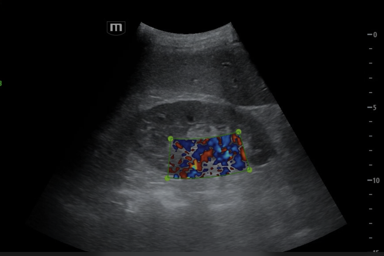
Figure 5: Color doppler flow placed on renal pelvis, negative for tinkling artifact.
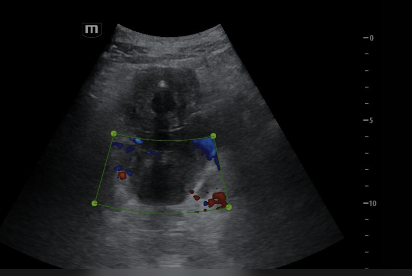
Figure 6: Color doppler placed near trigone of patient’s bladder without twinkling artifact or ureteral jets visualized.
Remaining ED course:
Patient had CT Abdomen and Pelvis that demonstrated bilateral hydroureteronephrosis with markedly enlarged prostate and an irregular-shaped fluid collection posterior to the bladder thought to represent a partially decompressed bladder diverticulum or other pelvic fluid collection. Additionally, a small right upper pole renal lesion was visualized with recommendation for further workup with outpatient MRI. Patient was given albuterol for his hyperkalemia and Rocephin for his urinary tract infection. Foley was placed for urinary retention. Nephrology was consulted for patient’s hydronephrosis and it was recommended that patient be given 100mg IV Lasix, 1L IV fluids run slowly, and a repeat renal panel after IV fluid administration. Repeat RFP demonstrated Na 129, K 4.5, and Cr 3.01. Patient was admitted to inpatient medicine for his bilateral hydronephrosis and AKI secondary to urinary outlet obstruction.
Brief hospital course:
During his admission, nephrology followed patient and urology was consulted. Patient had improvement of his obstructive uropathy with foley catheter placement. Patient’s AKI and hyperkalemia resolved with improvement of obstruction and IVF. Patient’s urine culture had no significant growth. Patient was discharged to SNF on hospital day 4 with remaining course of PO antibiotics, foley catheter in place, and plan to follow-up with urology outpatient for surgical intervention for his enlarged prostate.
Conclusion:
Bedside POCUS was used in the case of a patient presenting with altered mental status found to have AKI, hyperkalemia, and urinary retention. POCUS was performed prior to CT imaging and identified bilateral hydronephrosis, urinary retention, and enlarged prostate as cause of patient’s obstructive uropathy. This enabled the provider to gather critical information at bedside to expedite nephrology consultation and plan for hospital admission disposition before definitive imaging and final reads were obtained. This case demonstrates the bedside POCUS as an efficient and effective tool in advancing patient care in the emergency department.
References:
1. Deschamps J, Dinh V, Ahn J, et al. Renal ultrasound made easy: step-by-step guide. POCUS 101.Accessed October 23, 2026.https://www.pocus101.com/renal-ultrasound-made-easy-step-by-step-guide/
2. Deschamps J, Dinh V, Ahn J, et al. Bladder ultrasound made easy: step-by-step guide. POCUS101. Accessed October 24, 2026.https://www.pocus101.com/bladder-ultrasound-made-easy-step-by-step-guide/
Elena Ghezzi
Hydronephrosis
10/2026